Eye disease - Glaucoma (1 of 3]
Glaucoma facts
- Glaucoma is a disease that is often associated with elevated intraocular pressure, in which damage to the eye (optic) nerve can lead to loss of vision and even blindness.
- Glaucoma is the leading cause of irreversible blindness in the world.
- Glaucoma usually causes no symptoms early in its course, at which time it can only be diagnosed by regular eye examinations (screenings with the frequency of examination based on age and the presence of other risk factors).
- Intraocular pressure increases when either too much fluid is produced in the eye or the drainage or outflow channels (trabecular meshwork) of the eye become blocked.
- While anyone can get glaucoma, some people are at greater risk.
- The two main types of glaucoma are open-angle glaucoma, which has several variants and is a long duration (chronic) condition, and angle-closure glaucoma, which may be a sudden (acute) condition or a chronic disease.
- Damage to the optic nerve and impairment of vision from glaucoma are irreversible.
- Several painless tests that determine the intraocular pressure, the status of the optic nerve and drainage angle, and visual fields are used to diagnose glaucoma.
- Glaucoma is usually treated with eyedrops, although lasers and surgery can also be used. Most cases can be controlled well with these treatments, thereby preventing further loss of vision.
- Much research into the causes and treatment of glaucoma is being carried out throughout the world.
- Early diagnosis and treatment is the key to preserving sight in people with glaucoma.
What is glaucoma?
Glaucoma is a disease of the major nerve of vision, called the optic nerve. The optic nerve receives light-generated nerve impulses from the retina and transmits these to the brain, where we recognize those electrical signals as vision. Glaucoma is characterized by a particular pattern of progressive damage to the optic nerve that generally begins with a subtle loss of side vision (peripheral vision). If glaucoma is not diagnosed and treated, it can progress to loss of central visionand blindness.
Glaucoma is usually, but not always, associated with elevated pressure in the eye (intraocular pressure). Generally, it is this elevated eye pressure that leads to damage of the eye (optic) nerve. In some cases, glaucoma may occur in the presence of normal eye pressure. This form of glaucoma is believed to be caused by poor regulation of blood flow to the optic nerve.
How common is glaucoma?
Worldwide, glaucoma is the second-leading cause of irreversible blindness. In fact, as many as 6 million individuals are blind in both eyes from this disease. In the United States alone, according to one estimate, more than 3 million people have glaucoma. As many as half of the individuals with glaucoma may not know that they have the disease. The reason they are unaware is that glaucoma initially causes no symptoms, and the subsequent loss of side vision (peripheral vision) is usually not recognized.
What causes glaucoma?
Elevated pressure in the eye is the main factor leading to glaucomatous damage to the eye (optic) nerve. Glaucoma with normal intraocular pressure is discussed below in the section on the different types of glaucoma. The optic nerve, which is located in back of the eye, is the main visual nerve for the eye. This nerve transmits the images we see back to the brain for interpretation. The eye is firm and round, like a basketball. Its tone and shape are maintained by a pressure within the eye (the intraocular pressure), which normally ranges between 8 mm and 22 mm (millimeters) of mercury. When the pressure is too low, the eye becomes softer, while an elevated pressure causes the eye to become harder. The optic nerve is the most susceptible part of the eye to high pressure because the delicate fibers in this nerve are easily damaged.
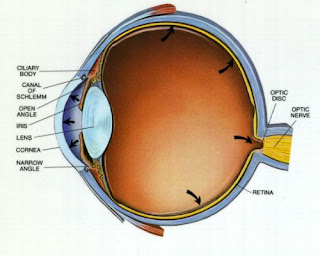
The front of the eye is filled with a clear fluid called the aqueous humor, which provides nourishment to the structures in the front of the eye. This fluid is produced constantly by the ciliary body, which surrounds the lens of the eye. The aqueous humor then flows through the pupil and leaves the eye through tiny channels called the trabecular meshwork. These channels are located at what is called the drainage angle of the eye. This angle is where the clear cornea, which covers the front of the eye, attaches to the base (root or periphery) of the iris, which is the colored part of the eye. The cornea covers the iris and the pupil, which are in front of the lens. The pupil is the small, round, black-appearing opening in the center of the iris. Light passes through the pupil, on through the lens, and to the retina at the back of the eye. Please see the figure, which is a diagram that shows the drainage angle of the eye.
Legend for figure: This diagram of the front part of the eye is in cross section to show the filtering, or drainage, angle. This angle is between the cornea and the iris, which join each other right where the drainage channels (trabecular meshwork) are located. The arrow shows the flow of the aqueous fluid from the ciliary body, through the pupil, and into the drainage channels. This figure is recreated fromUnderstanding and Treating Glaucoma, a human anatomy board book by Tim Peters and Company Inc., Gladstone N.J.
In most people, the drainage angles are wide open, but in some individuals, they can be narrow. For example, the usual angle is about 45 degrees, whereas a narrow angle is about 25 degrees or less. After exiting through the trabecular meshwork in the drainage angle, the aqueous fluid then drains into tiny blood vessels (capillaries) into the main bloodstream. The aqueous humor should not be confused with tears, which are produced by a gland outside of the eyeball itself.
This process of producing and removing the fluid from the eye is similar to that of a sink with the faucet always turned on, producing and draining the water. If the sink's drain becomes clogged, the water may overflow. If this sink were a closed system, as is the eye, and unable to overflow, the pressure in the sink would rise. Likewise, if the eye's trabecular meshwork becomes clogged or blocked, the intraocular pressure may become elevated. Also, if the sink's faucet is on too high, the water may overflow. Again, if this sink were a closed system, the pressure within the sink would increase. Likewise, if too much fluid is being produced within the eye, the intraocular pressure may become too high. In either event, since the eye is a closed system, if it cannot remove the increased fluid, the pressure builds up and optic-nerve damage may result.


No comments:
Post a Comment