Phimosis and Paraphimosis
- Author: Hina Z Ghory, MD; Chief Editor: Pamela L Dyne, MD
Background
Phimosis refers to the inability to retract the distal foreskin over the glans penis. Physiologic phimosis occurs naturally in newborn males.Pathologic phimosis defines an inability to retract the foreskin after it was previously retractible or after puberty, usually secondary to distal scarring of the foreskin.
Paraphimosis is the entrapment of a retracted foreskin behind the coronal sulcus. Paraphimosis is a disease of uncircumcised or partially circumcised males.
Pathophysiology
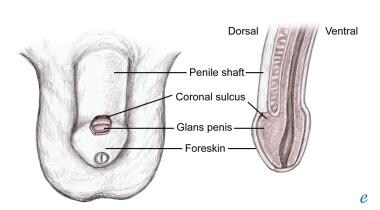
The uncircumcised male penis comprises the penile shaft, the glans penis, the coronal sulcus, and the foreskin/prepuce, as shown below.
Physiologic phimosis results from adhesions between the epithelial layers of the inner prepuce and glans. These adhesions spontaneously dissolve with intermittent foreskin retraction and erections, so that as males grow, physiologic phimosis resolves with age.
Poor hygiene and recurrent episodes ofbalanitis or balanoposthitis lead to scarring of preputial orifices, leading to pathologic phimosis. Forceful retraction of the foreskin leads to microtears at the preputial orifice that also leads to scarring and phimosis. Elderly persons are at risk of phimosis secondary to loss of skin elasticity and infrequent erections.
Patients with phimosis, both physiologic and pathologic, are at risk for developing paraphimosis when the foreskin is forcibly retracted past the glans and/or the patient or caretaker forgets to replace the foreskin after retraction. Penile piercings increase the risk of developing paraphimosis if pain and swelling prevent reduction of a retracted foreskin.
With time, impairment of venous and lymphatic flow to the glans leads to venous engorgement and worsening swelling. As the swelling progresses, arterial supply is compromised, leading to penile infarction/necrosis, gangrene, and eventually, autoamputation.
Epidemiology
Frequency
United States
Up to 10% of males will have physiologic phimosis at 3 years of age, and a larger percentage of children will have only partially retractible foreskins. One to five percent of males will have nonretractible foreskins by age 16 years.[1, 2]
History
Parents of patients with physiologic phimosis may bring in the patient after noting an inability to retract the foreskin during routine cleaning or bathing. Parents may also be alarmed by "ballooning" of the prepuce during urination — a normal finding.
Pathologic phimosis may be detected in males who report painful erections, hematuria, recurrent urinary tract infections, preputial pain, or a weakened urinary stream. (See below.)
Paraphimosis classically presents with a painful, swollen glans penis in the uncircumcised or partially circumcised patient. A preverbal infant may present only with irritability. Occasionally, the paraphimosis may be an incidental finding noted by a caretaker of a debilitated patient. (See below.)
Paraphimosis is classically seen in one of the following populations[3, 4]
- Children whose foreskins have been forcefully retracted or who forget to reduce their foreskin after voiding or bathing
- Adolescents or adults who present with paraphimosis in the setting of vigorous sexual activity [5]
- Men with chronic balanoposthitis
- Patients with indwelling catheters in whom caretakers forget to replace the foreskin after catheterization or cleaning
Urinary obstruction is a late feature.
Physical
Phimosis includes the following:
- The foreskin cannot be retracted proximally over the glans penis.
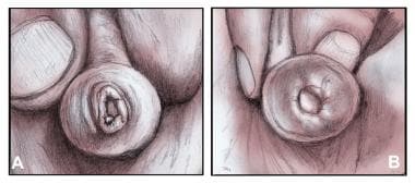
- In physiologic phimosis, the preputial orifice is unscarred and healthy appearing.
- In pathologic phimosis, a contracted white fibrous ring may be visible around the preputial orifice
Paraphimosis includes the following:
- The foreskin is retracted behind the glans penis and cannot be replaced to its normal position.
- The foreskin forms a tight, constricting ring around the glans.
- Flaccidity of the penile shaft proximal to the area of paraphimosis is seen (unless there is accompanying balanoposthitis or infection of the penis).
- With time, the glans becomes increasingly erythematous and edematous.
- The glans penis is initially its normal pink hue and soft to palpation. As necrosis develops, the color changes to blue or black and the glans becomes firm to palpation.


No comments:
Post a Comment